DIFFERENT STAGES IN THE EYE AND CORNEA AFTER BURNING BY BLISTERING AGENTS.
Click on the links to view the text and graphics
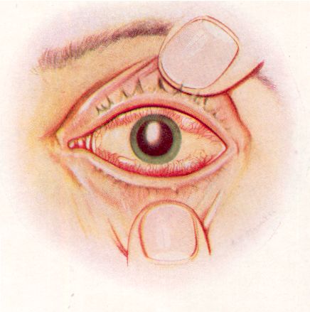
10. Severely Burned Eye in the Acute Stage
11 - Slightly later stage of acute burning of the eye (Plate 9)
12 - Stage of resolution after severe burning of the eye (Plate 10)
13 - Late stage of resolution (Plate 11)
14 - The cornea in the acute stage of severe burning (Plate 12)
15- The cornea - resolution after severe burning (Plate 13)
This correlates with the Cornea in Plate 12
(Plate 8)
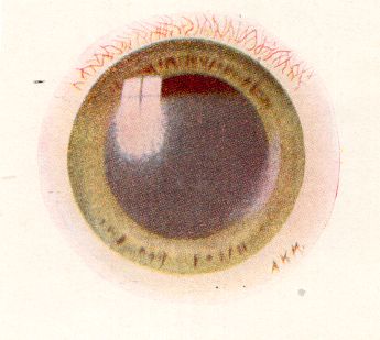
Relatively early after exposure to mustard gas vapour the eyelids and the external surface of the globe show an intense inflammatory reaction. Tears stream from between the closed oedematous eyelids, which may even be blistered, and there is often severe pain behind the eyes and in the forehead. The conjunctiva is swollen, oedematous, and bright red from injection of the blood vessels. The injury to the cornea, even when severe, is not so obvious, and careful examination is of great importance for its detection. Photophobia and blepharospasm render examination of the eye difficult.
The majority of gassed eyes exhibit inflammation of a general character that is not illustrated in this Atlas. But examples continually occur in which the eye is more severely burned, and these may be recognized by certain characteristic features that are depicted in the drawing. Whenever a dead white band crosses the exposed area of the conjunctiva, while the parts of this membrane covered by the upper and lower lids are red and oedematous, serious injury from the burning is likely to have occurred.
In the case illustrated, the caustic effect of the vapour is seen chiefly in the interpalpebral aperture. On each side of the cornea there is a dead white band due to coagulative oedema, which compresses the vessels and impairs the circulation, thus acting as a menace to the nutrition of the cornea. The swelling in the region of this white band is slight, while the protected conjunctiva above and below it is greatly swollen and injected and my even bulge between the lids.
The exposed portion is grey and hazy, it has lost its lustre, and when, viewed with a bright light and a magnifying glass it shows blurred "window reflex"' and a typical "orange skinned" surface. The haze gradually fades off above the region of the protected part of the cornea where the surface is usually bright and smooth. The pupil is at first contracted as the result of irritation and congestion. In this drawing it is shown as artificially dilated by atropine, which should always be used early in severe cases or where there is much pain and blepharospasm.
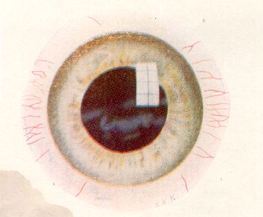
11. Slightly Later Stage of Acute Burning of the Eye
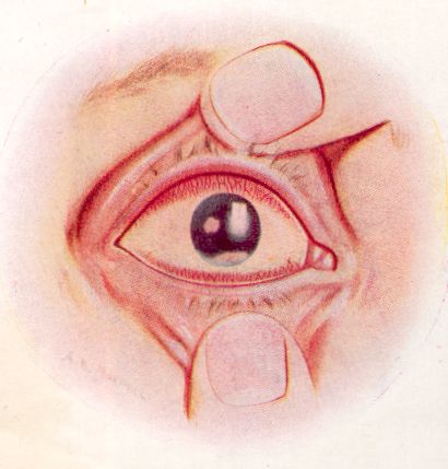
The swelling in the conjunctiva above and below has subsided, but the vascular injection remains, and the solid white oedema in the palpebral aperture is still well marked. The cornea is grey and lustreless in the exposed area.
With the lowering of the nutrition of the corneal epithelium, secondary infection is liable to take place. In this case an infiltrated corneal ulcer is seen associated with hypopyon. It is therefore important when there is conjunctival discharge, which indicates secondary infection, that in addition to the use of atropine the conjunctival sac should be cleansed by the instillation of antiseptic drops so as to check infection of any corneal ulceration which may develop. Otherwise the infective progress which has led to hypopyon may progress to panophthalmitis.
History of the case. The casualty was caused by a mustard gas shell bursting close to the man when he was riding a restive mule, and his box respirator was momentarily displaced. A fine spray of the liquid must have splashed lightly over his right side, for cutaneous blisters developed on this side only of his neck, cheek, and forehead. The right eye showed serious burning with the central white band, while the left eye was only in the state of general red conjunctivitis.
12. Stage of Resolution after Severe Burning of the Eye
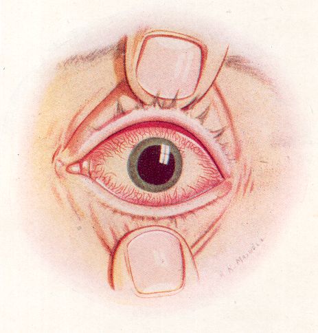
(Plate 10)
The vascular injection is passing off, the interpalpebral zone of solid oedema is becoming absorbed, and the corneal epithelium has regained its normal lustre. At this stage the use of atropine should be discontinued.
13. Late Stage of Resolution
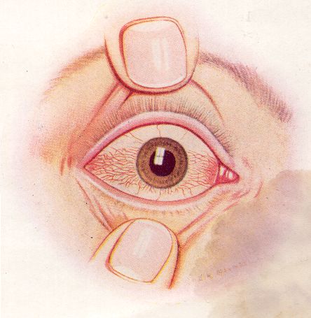
The earlier vascular injection above and below the cornea has practically disappeared; the solid white oedema has been absorbed, and the conjunctiva in the palpebral aperture now shows definite injection, often of a bright violet tint. The entire picture has changed, so that the parts which were red in the acute stage are now white, and the part which was formerly white is now red.
At this stage the use of atropine and shades should be abandoned. Astringent drops should be instilled and photophobia combated with cold douching, etc., while fresh air and occupation win help to restore the general health of the individual and mitigate any tendency to neurasthenia.
This drawing of the late phase of recovery after a severe burn would serve equally well to illustrate the early stages of a mild burn, which is the commonest form of eye lesion after exposure to the vapour rather than to droplets of the liquid. Though discomfort may
make the patient unable to open his eyelids, examination will in such mild cases reveal a lustrous cornea and a central band of injection instead of the central zone of white oedema that characterizes severe burns.
14. Drawing of the Cornea in the Acute Stage of Severe Burning
This corresponds with Plate 8. The exposed central area shows grey haze and loss of lustre on its stippled surface, which gradually fades off to the bright lustrous normal surface in the part above that has been protected by the eyelid. Injection of the conjunctival vessels is seen only in relation to this upper and less burned area.
15. Drawing of the Cornea in the Stage of Resolution after Severe Burning
(Plate 13)
The cornea is now smooth and bright with a clear light reflex on its surface. But some grey superficial nebulae are seen in the centre, and these may persist for several weeks. The injection of the conjunctival vessels is now limited to the central band.
16. Note on Late Changes
When the acute symptoms of a mustard gas lesion have subsided the vessels at the limbus in all but the mild cases are seen to show characteristic fusiform dilatations and varicosities
In the less severe cases these vessels do not show any tendency to invade the cornea to any extent, but in the more, severe lesions they tend to invade the superficial layers of the corneal stroma, and, where there is still active oedema of the cornea at the time when these vessels are growing in, an insidious, progressive vascularising keratitis is likely to develop and cause the case to drag on for years.
These cases of "delayed keratitis" are characterised by periods of relative freedom from symptoms, but unfortunately the corneal lesions tend to break down at intervals and to be very resistant to treatment.