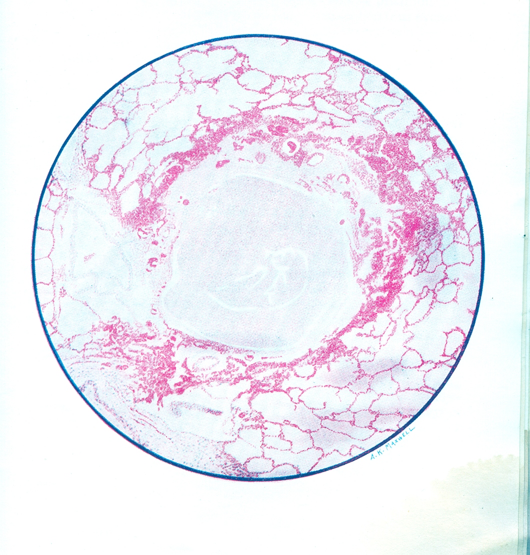
9. Microscopic Section of Human Lung after Mustard Gas Poisoning
Plate 7
Death occurred at the end of the second day after exposure (i.e., in 40 hours).
The bronchiole is filled with fibrin and pus cells, and its lining epithelium has been completely destroyed. The inflammation has caused a characteristic ring of haemorrhage in the tissues around the bronchial tube, and infection is beginning to appear in the alveoli nearest to these inflamed tissues. But there is no generalized pulmonary oedema and no disruptive emphysema.
Mustard gas may cause some catarrhal desquamation of the pulmonary endothelial cells, but it rarely excites an outpouring of oedema fluid from the pulmonary vessels. The pathological changes in the bronchioles and in the alveoli are therefore in the sharpest contrast with those caused by phosgene (see Plate I). As infection spreads into the lung tissues, patches of septic broncho-pneumonia and small abscesses develop, and these often excite an inflammatory oedema around them.
If the patient lives, his bronchial mucous membrane is slowly regenerated; and during this time he is naturally subject to reflex spasms of coughing or even to a protracted bronchitis.