
CHAPTER 7
BONY LESIONS OF THE DIAPHYSES
There is no traumatism that brings into such strong relief the striking differences separating war surgery from ordinary surgery as the bony lesions of the diaphyses. A surgeon who considers he can base their treatment on his general ideas would be liable, unfortunately for the wounded, to remain very inferior to the ideal of the duty he has undertaken. The adoption of the new projectiles has brought with it no important modification in the data derived from experiments on the dead body, on the one hand, and from experience acquired in recent wars on the other. According to world-wide statistics, lesions of the bones are seen during a campaign in a proportion of one-fifth of all wounds.
Cold-steel weapons lead to sections of the diaphyses; projectiles to contusions, cracks, and fissures, to fractures by contact, perforation of one side of the bone, perforation right through, and to grooves.
This classification should be adopted by all surgeons, in the first place because it is based on strictly exact and constant provisions, in the second place because it originates from our chief practical data.
LESIONS CAUSED BY PROJECTILES.
Contusions.--These are either the result of direct shock or of tangential contact of the projectiles. They are very frequently produced by bullets, but are often unperceived on account of their giving rise to no immediate signs. At the seat of the contusion the periosteum is involved and destroyed, and the marrow may or may not show either circumscribed or extensive pouring out of blood.
Cracks and Fissures. Isolated cracks and fissures of the diaphyses are also frequent, but, like bony contusions, they nearly always are unperceived. Wounded men who present long fissures of the bones of the lower limbs can walk when left to themselves.

A crack is a cleft whose sides are very near together, and can hardly be seen; a fissure is a visible cleft whose sides are widely separated.
The most remarkable are the longitudinal cracks and fissures, often very extensive; they are either single or multiple, but some are oblique, some curved. Isolated cracks and fissures are outlines of those that fix the limits of fractures by contact, of which they show the direction and position (Delorme).
Symmetrical fissure and opposite fissure are the most striking and the most constant lesions of the diaphyses. The former furrows the side of the bone that has not been hit, and this happens in the plane passing through the point of contact of the projectile. It is the result of a tangential contact (o.f.).
These fissures are seen on all the long bones. Absolute diagnosis up to now has been very difficult, for neither in bone contusions or in fissures was there anything characteristic with regard to shock, inability to use the limb, denudation of the bone, deviation in the course of the projectile, its change of shape if it remained in the limb, increase in size of the aperture of exit, relationship of the track to the bone.
Radiography sometimes affords a certain diagnosis, but this is not constant.
FRACTURES BY CONTACT. - These are the result of the direct or indirect (tangential) contact of a bullet fired point-blank, or that has ricocheted or been deflected.
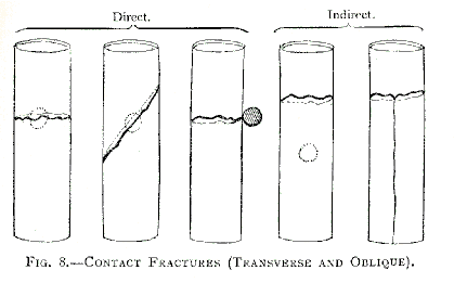
We recognise fractures by contact as transverse, oblique - that is to say, forming a simple line of fracture; and fractures that have large splinters of bone
TRANSVERSE AND OBLIQUE FRACTURES.--Relying on our experience, we have asserted that transverse and oblique fractures are not very rare. The teachings of the Transvaal War have confirmed our dictum, and the present war strengthens it still more.
Sometimes the fracture corresponds exactly to the point of the bone hit by the bullet (direct fracture), sometimes it occurs at 2, 3, 10 centimetres from the place of the bullet's contact (indirect fracture). Indirect fracture can be single or double, and its line can either be simple or accompanied by longitudinal fissures.
FRACTURES WITH LARGE SPLINTERS. - In these fractures, which are well known ever since we fully described them, the lesions extend more in the direction of the axis of the bone than perpendicularly to it.
Five different types will be found in the illustration, fig. 9.
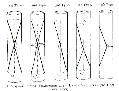
First Type: This is the most important. From the point of impact extend four diverging curvilinear fissures, the convexity of which is turned towards the axis of the diaphysis; these fissures are united on the side of bone which is not hit to the large symmetrical longitudinal fissure (s.f.) These fissures enclose two large triangular splinters facing one another at their apex; they are placed between the superior and inferior fragments; their pointed extremities have undergone no loss of substance. This is a point to remember.
These splinters may occupy a quarter, a third, or one half of the bone.
At the point of contact the periosteum is destroyed, the bone contused; all along the fissures it is raised by blood and marrow reduced to pulp; suffusion of blood is seen in the medulla
Second Type: This is a fracture with large splinters subdivided transversely or obliquely at their centre. (Rare.)
Third Type: Oblique longitudinal spiroid fracture. On the side of the bone corresponding to the point of contact there is a very oblique line in the form of an elongated S, which by its curved extremities joins the longitudinal symmetrical fissure. This exceptional type is almost exclusively seen in the femur (upper third).
Fourth Type: Fracture having the shape of a V, cuneiform and with one large splinter. It is the fracture of the first type, or in the form of an X, in which the line of one of the splinters is wanting. Of the two fragments one has the shape of a V, or of a wedge, and is sometimes above, sometimes below; the other takes the form of a radish. This fracture is frequent.
Fifth Type: Fracture with one splinter, and transverse subdivision of the remainder of the bone. (Rare.)
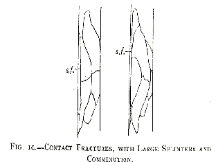
Such are the simple types, without comminution that are produced on the diaphyses by contact with bullets. The bone is fissured or broken; the fragments, whose limits are marked, are either held closely together by their serrated edges (in which case the continuity of the limb will not be interfered with so long as the bone is not subjected to any shock or to untimely exploration), or else the fragments and the splinters are from the very beginning separated as in an ordinary fracture.
The periosteum, which has become separated from the bone, is raised to the level of each secondary fissural tract; but whether the subdivided splinters remain in situ or are displaced by the faulty position of the superior or inferior fragments, the comminuted fracture by contact retains its main characteristics: the extremity of its fragments is SHARP, WITHOUT LOSS OF SUBSTANCE; THERE IS NO FREE SPLINTER; all the splinters are adherent.
The conditions governing the manner of arrangement of fractures by contact with large splinters give the keynote to lesions connected with perforations or grooves, for, as we have proved, a perforation or a groove is but a fracture by contact with a perforation or a groove superadded.
Perforations.--By experiments we have proved that fracture caused by perforation, whether with or without comminution, is the habitual form of fracture produced by firearms.
We have recognised two kinds which have been generally accepted: - Incomplete perforations -- that is to say, perforations of only one side of the bone -- and complete perforations when both sides of the diaphysis are involved.
Incomplete perforations. -- that is to say, perforations of only one side of the bone -- can only be produced (and this is easily understood) by bullets whose velocity cannot be very great, since they have not been able to continue their course. Therefore, as comminution of a fracture is in inverse ratio to the extent of motion acquired by the bullet, it is evident that these fractures must always be simple in type. On the other hand, however, we must not forget that the bullet has not only made a perforation in the diaphysis, but that, by its simple contact, before causing the perforation, it has given rise to the longitudinal fissures seen in fractures by contact.
It is the first X type of fractures by contact that is connected with perforation of only one side of the bone. With the fissures and the large splinters, which we need not describe again, there is an orifice in the bone, generally rounded in shape, sometimes oval whose diametrical dimensions are less than those of the bullet. The latter may be in the medullary canal at the level of its aperture of entry; occasionally it has slipped down the canal, or it may have come into contact with the inside of the opposite wall of the bone, giving rise to some short splinters, but as it has not sufficient strength left to emerge, it remains where it is.
COMPLETE PERFORATIONS. -The projectile that produces them having a sufficient but variable active power to go through the two bony walls, complete perforations are either more or less simple, or else comminuted even excessively comminuted.
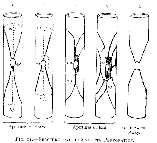
In regard to the general direction of the fissures, the limitation of the splinters and the shape of the fragments, fracture by complete perforation nearly always shows much resemblance to fracture by contact with two more or less subdivided large lateral splinters, or to cuneiform V-shaped fractures (Fig. 11, 1 and 2).
The bony aperture of entry is circular and regular, of the same dimensions as the projectile, or smaller, occasionally oval in shape. The bony aperture of exit is variable in form. Rarely circular and regular, it is nearly always, owing to loss of substance, more or less quadrilateral, its borders being formed by the splinters that the projectile has detached (fig. 11, 3 and 4). These splinters, more or less free, are then stationary; they can, however, be thrown off.
In such an instance the bullet has not acted alone, as in a contact fracture or in perforation of only one bony wall. The splinters it has torn off at the aperture of entry, the subdivided splinters close to the track to which the bullet has communicated part of its active power, the fragments of the bullet which may have broken in pieces, have all acted as secondary projectiles. These last, propelled towards the bony aperture of exit in a more or less irregular manner, have increased the damage, and have given rise in some cases to a fracture feeling like a bag of nuts; even in some instances the seat of fracture has been freed from splinters (focus swept away, bullet fired from a very short distance-- Fig. 11, 5)
Grooves.--In frequency they come after perforations.
They are more or less deep furrows in which one might accommodate a quarter, a half, even three-quarters of the diameter of the bullet; if deeper, the groove would become a perforation.
Grooves generally affect but a very small extent the transverse diameter of the bone. We must differentiate the grooves of the ridges from those of the body of the diaphyses.
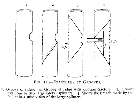
Grooves of the Sides and of the Ridges of the Bone. When the anterior ridge of the tibia, the sides of the same bone, the edges of the inferior extremity of the humerus, the linea aspera, the sharp edges of the radius or of the ulna, are indented, the indentation is sometimes distinct and isolated, sometimes it is accompanied by a transverse or an oblique fracture (Fig. 12)
Grooves on the Body of the Diaphyses.-- They have a close relationship to the types of contact fractures we have described, especially to the first and the fourth (type with large splinters, the cuneiform V-shaped type).
As the bone, in the tangential contact that precedes the abrasion causing a groove, cannot have received a shock from active force as intense as the one it would experience when struck point-blank, which would lead to perforation, the groove may generally be placed in the type in which there is little or but slight comminution. This is an important fact.
It results from this that a great many grooves exist without any solution of continuity of the bone. Undoubtedly we may see rather shallow grooves of the diaphyses which consist of abrasions without fissures, but this is exceptional, and usually a fracture by contact accompanies the grooving of the bone.
When the bullet has bored in the body of the bone a rather deep track, it has not only indicated the limits of one or two large splinters, the lines of whose fissures join the big fissure of the wall perpendicular to the track of the bullet (opposite fissure) but also it has brought about, symmetrically at its point, a short symmetrical longitudinal fissure, and in the centre of this subdivision of splinters, in the centre of this secondary splinter, which to all intents and purposes is adherent, the bullet has travelled, piercing a narrow track in its passage.
In complete perforations the shape of the bullet is altered; it subdivides if it is provided with a covering. As we have before pointed out, it changes many of the splinters into secondary projectiles, which increase in number, and by their comminution extend the field of damage. In such a case there is no change of shape of the bullet, the reduction of the splinters into fragments is shown by their transformation into dust, which is gently projected into the soft parts.
Fracture by groove can be distinguished from the multiplicity of fractures through perforation by its invariable simplicity
SPLINTERS are pieces of bone whose limits depend on the projectile; they are connected with the foci of the fractures, and may be divided into free and adherent.
1. FREE SPLINTERS.--The smallest of these may only be represented by a kind of glazed bone-dust, resulting from the first hard, bony layer met with by the projectile. They are found lying around the aperture of entry into the bone.
Most free splinters come from the bony aperture of exit. With them are sometimes found splinters that have been set free from the lateral walls where large splinters have been subdivided.
These free splinters, therefore, cannot exist in contact fractures or in incomplete perforations. This is an important dictum which surgeons should bear in mind.
The higher the velocity of the projectile causing them, the shorter they are. In the great majority of cases they correspond, as we have before remarked, to the MUSCULO-CUTANEOUS CANAL of exit in which the bullet has left them.
We must not forget that this musculo-cutaneous canal extends from the aperture of exit in the bone to the aperture of exit in the skin. It would be useless, and also incomprehensible, to search for these free splinters anywhere else in cases that have been wounded by ordinary rifle-fire.
The greater the velocity of the projectile, and also the more the fracture shows comminution, the farther these splinters will be from the aperture of exit in the bone.
When the velocity is excessive, free splinters are no longer carried along, but are violently thrown out in the form of a sheaf. They no longer have any exact situation, they bury themselves in the soft parts at a more or less long distance from the track, and some of them break out of the limb through numerous separate orifices.
2. ADHERENT SPLINTERS. These correspond to the parts of the bony cylinder that the projectile has not touched.
Free splinters are short, adherent splinters are 4, 6, 8, l0, and even 20 centimetres long.
A large splinter is never a free splinter, but always an adherent splinter.
Their dimensions, like their number, are inversely in proportion to the velocity of the projectile. The less the velocity, the larger the adherent splinters and the less their number, and inversely.
The firmness of their adhesions also depends on the velocity of the bullet. The less the latter, the firmer the former.
The size and extent of these splinters are in close relationship to the wounded bone. On the femur and the tibia they are often enormous; they decrease in size on the humerus, the clavicle, the bones of the forearm, and the metacarpal and metatarsal bones.
The direction taken by the bullet has also, in this connection, a certain importance. A bullet, the direction of which is nearly that of the axis of the bone, or follows it (enfilading fire), gives rise to longer adherent splinters than one that strikes perpendicularly.
The Osseous Focus.- from what has already been said, it is seen that the osseous focus of fracture by firearms presents, in the large majority of cases, two fragmentary extremities moulded into the form of a wedge: a SHARP wedge (contact fractures), a BLUNTED wedge (perforations); also invariably adherent splinters (all fractures), often free splinters (perforations).
Splinters increase the size of the focus by 4 to 20 centimetres, habitually by 6 to 8 centimetres. Only in a cleared- out focus (excessive velocity) is there loss of substance, and even then it is very trifling, because it is entirely derived from the splinters. The length of the bone is not obviously diminished. Even in these cases the total loss of substance, especially when considered from the point of view of the two cuneiform fragments, the upper and the lower does not exceed, in spite of appearances, 2 centimetres.
Notwithstanding this damage done to the bone, even when there is a great deal of comminution, the soft parts have suffered the more injury; but we know how easily they undergo repair.
As a general rule, the higher the velocity of the projectile, the more limited in length is the fracture, but at the same time it is more comminuted.
This is a most important fact which we include amongst the many others we have stated. On the battlefield it affords a key to the nature, the importance, the intricacy of the assistance we have to render in cases of fracture.
The focus shows a difference according as the bone has not sustained a solution of continuity, or, on the contrary, according as its continuity is interrupted. It also shows a difference according as:
(1) the focus is simple, without splinters;
(2) or simple with stationary splinters that have not been displaced;
(3) or simple with splinters that have been displaced and thrown out.
In the first case the canal of exit in the soft parts is narrow, it turns on itself and has no tendency to infection; in the second case it is widely open much contused and bleeding; in the third case it may reach the dimensions of the thumb and even more, it may admit several fingers held close together, even the whole hand. It is much inflamed.
If the focus of a fracture by firearms is comminuted, this does not indicate that there is any solution of continuity of the bone itself. In this connection fractures caused by firearms differ entirely from the fractures seen in ordinary practice in which comminution is always accompanied by solution of continuity.
DIAGNOSIS OF OSSEOUS LESIONS OF THE DIAPHYSES.
The diagnosis should rest on two points:
(1) General diagnosis of the osseous lesion;
(2) diagnosis of the group and of the variety.
1. GENERAL DIAGNOSIS.-- Pain, loss of all power in the limb, change in its length or in its shape, abnormal mobility, all these points can be of use to us in fractures by firearms, as they are in ordinary fractures, in establishing a general diagnosis; but as many of these signs very often are wanting, we are obliged to look for others. The following are the signs we consider of the greatest importance. They are:
Shock (comminuted and very comminuted fractures).
Pain evoked by pressure at a distance or on the supposed line of the fissures.
Angular prominence of the terminal extremity of the large splinters, easily felt in the superficial foci (tibia, ulna, clavicle), and sometimes in bones situated more deeply, even in the femur.
Position of the wounds in direct relation to the superficial bones (hand, foot, tibia, ulna, clavicle).
The relation of the track in the soft parts to the position of the bones.
The enlarged dimensions of the aperture of exit in the skin and in the clothing compared with the aperture of entry (short and middle range firing).
The spread-out form of certain orifices made by soft lead bullets.
Swelling profuse haemorrhage (the bone becoming a regular enormous collection of blood). This sign has not been sufficiently dwelt upon.
Escape of small oily drops (comminuted fractures of the big long bones).
The presence of free splinters in the canal of exit, at the level of the cutaneous orifice, or that of the clothes. This is a favourable sign.
A special change of shape of the projectile (lateral change of shape, bending back of the point), even when the aperture of entry shows from its appearance and its dimensions that the bullet has entered from point-blank firing, and had not been deflected nor had its shape altered.
Extensive crepitation which is obtained by bringing the splinters together; or localised crepitation, obtained by slight compression exercised in the direction of the aperture of exit in the bone. These are quite harmless proceedings, very different to the highly reprehensible plan of seeking for crepitation by moving the whole of the bone, or by rotating the fragments; this, indeed, is still worse, for this rotation easily renders complete an incomplete fracture, and gives rise to displacements which are difficult to correct.
These, then, were the signs we brought forward. They well maintain their value, and very often the military surgeon is unable to obtain others. Under favourable conditions, at the rear, radiography, the generalisation of which becomes more and more necessary, much simplifies nowadays the general diagnosis.
When in doubt, we should act as if the fracture existed, and a more or less rapid examination, or one carried out subsequently, will either confirm or nullify the diagnosis.
2. DIAGNOSIS OF THE GROUP AND OF THE VARIETY--
(I) Contact Fractures.--We have given as principal signs of these fractures: absence of aperture of exit, absence of very small oily drops, absence of free splinters in the canal of exit, and, the best sign of all, absence of perforation of the bone or of indentation, this having been proved by direct exploration.
Here radiography has furnished precise indications and simplified research after these last two valuable signs. In fact, radiography has completed the clinical history of this group.
It shows in these contact fractures with large splinters-- The absence of splinters in the canal of exit, and, above all, the PATHOGNOMONIC SIGN: the SHARP WEDGE OF THE TWO FRAGMENTS, upper and lower. In no other kind of fracture caused by firearms is this sign to be found.
(2) Fractures by perforation.-- Radiography settles the diagnosis of fractures by perforation of one wall of the bone.
Fractures by perforation of the two walls of the bone are recognised by the rectilinear track in the axis of the bone, by the enlargement of the aperture of exit in the soft parts and in the clothes, by the presence of free splinters close to the cutaneous aperture of exit or else in the track of exit, by multiple orifices (explosive fire), by the change in shape of the point of the bullet, by splitting up of those bullets that have an envelope, by the localised crepitation in the focus of free splinters near the aperture of exit in the bone.
Thanks to these signs, the diagnosis of the lesion is generally easy. Radiography has made it still easier by disclosing (1) when there is no solution of continuity, the ROUNDED OR OVAL PERFORATION the diaphysis has sustained in the first wall that has been pierced, the more irregular but as easily demonstrated loss of substance in the second wall; (2) when there is solution of continuity, and even considerable displacement of the fragments, the INDENTATION presented by the superior and inferior cuneiform fragments; finally (3) in both cases the PRESENCE OF NUMEROUS FREE SPLINTERS, either lying in the canal of exit or moved into a new position.
(3) Fracture by Groove.--These fractures were very difficult to diagnose before the advent of radiography. The circular nature of the track, occasionally the slight change of shape of the bullet (lateral parts and apex), the small free splinters in the canal of the wound and especially the verification by the finger of a peripheric osseous groove, were the signs met with.
Radiography renders the FOLLOWING PATHOGNOMONIC SIGN perfectly clear: PERIPHERIC INDENTATION in the osseous track of hard lead bullets with an envelope (German and Austrian bullets). These tracks are rendered evident by small seed-like particles of lead when the bullet has become separated from its covering.
Comminution is easily recognised. It is shown--(1) By multiplied loud, fine crepitation of free splinters, very different with regard to sensation and to sound from the extensive crepitation, more muffled and not multiplied, caused by the friction of the long adherent splinters. (2) By the presence of a large number of splinters. We must also remember that in war surgery grave comminution and a solution of continuity are not synonymous.
Not only has radiography thrown light on the general diagnosis of these fractures, and allowed us to establish the diagnosis of the different groups, but every day it enables us to identify metallic foreign bodies, whole bullets that have lost their shape or become subdivided, and have been arrested in the osseous focus or in the neighbouring soft parts after having caused the fracture of the diaphysis.
We have already described many of these changes of shape, but in doing so we always had before our eyes the changes of shape that result from contact with hard soil before reaching the human body. Now we have to deal only with those that result from contact with bone.
Changes of Shape in Bullets that have struck Bones. - 1. Soft lead bullets that have caused FRACTURES BY CONTACT are flattened out, and often take on the shape of the bones they have struck. According to the bone it has reached, the bullet is flattened or concave.
2. It is the same thing with hardened lead bullets that have an envelope. The change of shape consists especially in flattening of the apex, with or without separation from the envelope; but in these cases, again, the surface is flat or concave.
3. With bullets composed of one piece, such as the D bullet, the change of shape is insignificant. In PERFORATIONS, both soft lead bullets and hardened lead bullets with an envelope become flattened are compressed, and become bent from the apex to the base. The flattened surface of the apex is rendered irregular. The increase of diameter, consequent on the compression, results in enlargement of the bony aperture of exit, in the liberation of more splinters, and also in enlargement of the aperture of exit in the soft parts.
Though less marked, the changes of shape in the D bullet are analogous, but do not present any notable irregularity in the surface of the turned-back apex.
With GROOVES, the changes in the shape of the bullets are insignificant.
Foreign Bodies derived from the Clothes.--Diagnosis of foreign bodies derived from the clothes is rendered certain by inspection of the clothes which at the aperture of entry show loss of substance, and indicate the number and the dimensions of the pads, consisting of pieces of clothing, that are in the wound. The surgeon should never forget to make this examination. The enlarged aspect of the aperture of entry WILL ALONE determine the probability of the sojourn of these infecting bodies in the focus; on the other hand, increase in size of the apertures of exit is a sign that makes us presume the existence of a lesion of bone.