 WWI Resource CentreWWI Resource Centre
Here are some scans of the effects of mustard gas from the official Medical Manual of Chemical Warfare, 1940, based on data from 1918; the acknowledgement to go to HMSO.
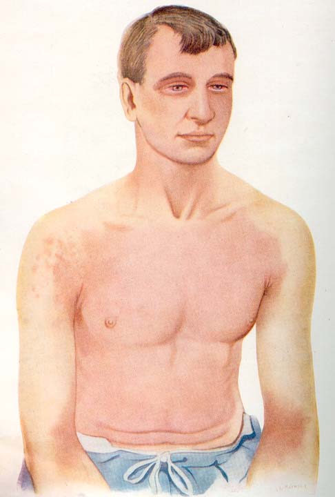
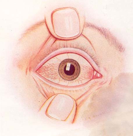
Left is a drawing of a soldier who was exposed to mustard gas at Ypres in 1917. He wore a box respirator for only 30 minutes and he was exposed without any protection for four hours, luckily the gas concentration was only slight. He developed conjunctivitis and vomiting some hours later and this drawing was made on the fifth day. It shows reddening of the skin and conjunctivitis.
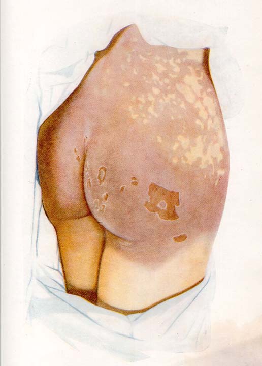
Right is a drawing from a man who sat on the ground contaminated by mustard. He burnt his buttocks and scrotum and the mustard initially caused a reddening, similar to skin1.jpg within 24 hours, but then he developed blisters, aggravated by pressure. On the eighth day the redness changed to a brown staining. This drawing was made on the eleventh day. Infection was avoided and he was healed by three weeks.
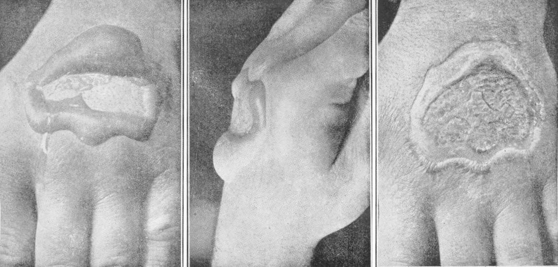
This series of photos are of a man whose leather glove was accidentally contaminated on its back by mustard. Photos 1 and 2 were taken one day and photo 3 one week later.
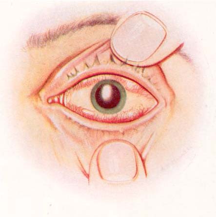
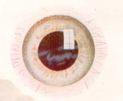
Eye A Cornea A Eye B The eye pictures are of a man who was riding a mule and a mustard gas shell burst close by; the mule bucked and the man's respirator was momentarily displaced, allowing a fine spray of mustard to affect his right side of face. "Eye A" is a drawing of the right eye which was severely burnt. "Cornea A" is a close up of the cornea, the transparent part of the eye. The pupil has been dilated by atropine as part of his treatment.
"Eye A" shows inflammation and swelling of the conjunctiva, the skin overlying the eye and "Cornea A" demonstrates opacity of the cornea below the upper part that has been protected by the eyelid.
"Eye B" shows a slightly later stage than A. The swelling of the conjunctiva has settled but the congestion remains and there is pus in the anterior chamber of the eye, associated with a corneal ulcer. Much pain would be associated with the ulcer and the prognosis has to be guarded for loss of sight.
Eye C Cornea B Eye D "Eye C" is the stage of resolution of this man who fortunately recovered. Atropine was discontinued and "Cornea B" shows that the transparency of the cornea is returning. The grey markings persisted for several weeks. Eye covering to exclude light has to be continued however.
"Eye D" shows a later stage which can be expected to recover. The conjunctivitis is settling and is no longer general, now being localised to the sides of the eye. Atropine had been stopped and the eye shade can be discontinued.The cornea is almost normal.
submitted: August 23, 2004 by Geoffrey Miller